By Dave Palmlund
For an authority on particle analysis who’d earned an award for a peer-reviewed paper on visual inspection standards of injectable drug products, it was disconcerting. Using imaging particle analysis, a technology available to industrial markets for less than a decade, Berdovich’s efforts to solve his customers’ problems led to his own business success.
His company, Wheeling, Ill.–based contract laboratory Micro Measurement Laboratories, Inc., does particulate matter testing, particle identification, method validation and visual inspection standards development for pharmaceutical and biotechnology companies worldwide, among other services.
The company offers particular expertise in standards and recommendations set by the U.S. Pharmacopeial Convention (USP), including USP <787>, which covers the testing of sub-visible particulate matter in therapeutic protein injections; USP <788>, which covers testing for and reporting of sub-visible particulate matter in injections and parenteral infusions; and the forthcoming USP <790>, which covers the inspection of visible particulates in injectable drug products; along with the revisions USP <1787>, <1788> and <1790>.
"I was getting very frustrated that when it came to sub-visible or semi-transparent particles like proteins, we were essentially in the dark," says Berdovich, "and USP <788> is our bread and butter."
That was three years ago and the fact that everyone else operated in the same darkness offered little comfort.
Why it hurt
Berdovich’s team used light obscuration instruments to detect particles, as prescribed in USP <788>. The problem, he realized, was that light typically travels through the particles without being scattered. Proteins and protein aggregates, along with other translucent and transparent particles, often defy detection by even the finest light obscuration devices, especially when in the sub-visible range of less than 20 µm. Additionally, much larger particles are often severely undersized when measured, if detected at all.
Detecting these particles is even more challenging using manual visual inspection — looking at individual vials under controlled lighting conditions and determining whether visible particles are present using the naked eye. This method has limitations similar to those using light obscuration, and because it is inherently subjective, it can yield unreliable results, especially in some biologics and lyophilized drugs.
At present, 100% visual inspection is required for visible particulates and two methods, light obscuration and membrane microscopy, are accepted by USP to test for the presence of sub-visible particles in parenteral formulations.
Since most manufacturers aim primarily for compliance, there is little incentive to learn more about the formation or introduction of particles present in compliant formulations. For example, under USP <788>, sub-visible particle testing is typically done in a lab away from the line where visual inspections are conducted for visible particles. Vials may be rejected from either test but are not often evaluated together using orthogonal methods to determine what particles are triggering the rejections. Concern is focused on effectively rejecting off-spec vials rather than on identifying the type of particle detected and understanding why it is present.
The situation faced
According to Berdovich, this mindset is changing. "Most of my customers want to know more about their products than compliance requires, such as particle shape — it’s invaluable in case problems arise later in formulation — but you can’t find out much more if you’re using laser scatter or light obscuration devices," he says.
The problem came to a head during another test that required measuring particles in a liquid or gel formed on a membrane. Since the particle shape deformed in minutes, the analysis needed to be done quickly. Manual microscopy proved far too slow. Moreover, the particles deformed when filtered and squeezed between the glass slides. "We were bumping up against a wall and thought there was no way to do this," says Berdovich.
Then he read in a trade magazine about new instrumentation: the FlowCAM, from Fluid Imaging Technologies, Yarmouth, Maine.
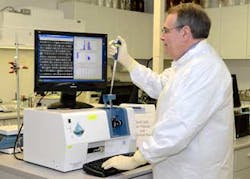
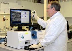
The FlowCAM imaging particle analysis system automatically detects the presence of particles and microorganisms in a sample, including transparent, semi-transparent and sub-visible particles from 2 µm to 800 µm in size.
The particle analysis instrument, using digital imaging technology, was first developed for use by oceanographers. Since 2004, it has found use in industrial markets including food & beverage, chemicals, abrasives and plastics, as well as pharmaceutical.
The instrument today includes cross-polarized illumination, laser and fluorescence-triggered acquisition and use of an optional high-precision syringe pump. The instrument’s software package does typical spreadsheet analysis operations on particle data while presenting the results visually as images — as opposed to tabular representation. It incorporates statistical pattern recognition for particle characterization, a big improvement over simple value filtering.
Not relying on light scatter
Rather than rely on light scatter, the FlowCAM’s imaging technology captures a high-resolution, digital image of each individual particle passing in front of its flow cell. Imaging thousands of particles in seconds, it measures each one based on its actual image to yield data based on particle shape, size and transparency.
Clicking an image or touching the image on screen reveals the measurement data, which may be graphed several different ways depending on how the data needs to be visualized and the types of particles targeted.
More than 30 different properties, including length, width, diameter, volume and aspect ratio, as well as advanced morphological types such as circle fit, elongation, perimeter and roughness are measured. Fluorescence and color are also available as options. Each image is saved with its corresponding measurement data for review, analysis and sharing online or by email.
Fluid Imaging Technologies customers include the U.S. Food and Drug Administration, Pfizer, Amgen, Roche, Nestle and the U.S. Environmental Protection Agency.
"You can get more information from the FlowCAM," says Berdovich.
From the advent
Now that he and his analytical staff have more than three years of experience running analyses on the instrument, he says, "Going to the FlowCAM with a particle problem is just the best feeling in the world because it turns data into useful information a customer can use to solve a problem."
For one application, FlowCAM measures the coverage of coatings on medical devices. If the coating process is insufficient, particles may be released that could affect the patient. "These types of particles simply can’t be effectively characterized by anything but a FlowCAM," says Berdovich.
Similarly, the FlowCAM may be used to optimize microencapsulation processes by looking at the shell layer around individual particles.
The ability to automatically differentiate one type particle from another, such as a globular protein aggregate from a curly fiber, round oil droplet or air bubble, also proves useful. Foreign matter, such as silicone oil droplets, for example, may come into contact with a sample from a rubber stopper. To a light obscuration device, these opaque oil droplets are counted just as if they were a protein or any other particle, since no distinction from one to the other may be made. This artificially increases the total particle count and may trigger destruction of an entire batch of quality product for failing to meet specifications.
While microscopic membrane analysis can verify that these droplets are not discrete, it cannot sufficiently characterize their concentration, since they pass through the membrane. This can be troublesome because the concentration is typically needed to solve a problem.
To the outcome
On the other hand, the FlowCAM images each individual oil droplet and the total count of droplets — or of any other type of particle — may be characterized as an isolated population, or even removed from the total particle count. "Again, you just can’t do this with anything but a FlowCAM. The data tells a lot," Berdovich adds.
The lab has provided several customers with FlowCAM data for submission to the FDA. "When you’re submitting data to the FDA, you want to understand the issues and you don’t want any doubt about the accuracy," Berdovich says. "I feel very confident in the FlowCAM because it helps us look under every rock when solving a problem, and it has also opened a lot of doors for us."
Since adding the FlowCAM, Berdovich has substantially increased his laboratory services customer base among medical device and pharmaceutical product manufacturers. Growth triggered a facility expansion, with an added clean room and staff. "With the FlowCAM and our knowledge base, we can take on applications that other labs won’t,” says Berdovich.
Fluid Imaging Technologies today provides particle services to more than 20 other laboratories. "Now that we have the ability to see exactly what’s in a sample, there really isn’t any excuse to look only as far as the USP requires."
Dan Berdovich is the owner of Micro Measurement Laboratories, Inc. He may be reached at (847) 459-6540, [email protected]. Dave Palmlund is sales manager for Fluid Imaging Technologies. He may be reached at (207) 289-3200, [email protected].

